
Donkey Secondary Antibodies


Biotium PTH / Parathyroid Hormone (N-Terminal)(PTH/1175), CF647 conjugate, 0.1mg/mL
The epitope of this MAb maps in the C-terminus of PTH, a hormone produced by the parathyroid gland that regulates the concentration of calcium and phosphorus in extracellular fluid. This hormone elevates blood calcium levels by dissolving the salts in bone and preventing their renal excretion.It is produced in the parathyroid gland as an 84 amino acid single chain polypeptide. It can also be secreted as N-terminal truncated fragments or C-terminal fragments after intracellular degradation, as in case of hypercalcemia. Defects in this gene are a cause of familial isolated hypoparathyroidism (FIH); also called autosomal dominant hypoparathyroidism or autosomal dominant hypocalcemia. FIH is characterized by hypocalcemia and hyperphosphatemia due to inadequate secretion of parathyroid hormone. Symptoms are seizures, tetany and cramps. FIH exist both as autosomal dominant and recessive forms of hypoparathyroidism.Primary antibodies are available purified, or with a selection of fluores

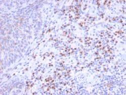
Biotium RCAS1 / Estrogen Receptor Binding Site Associated, Antigen 9(CPTC-EBAG9-1), CF647 conjugate, 0.1mg/mL
EBAG9, also known as RCAS1, is an estrogen-transcribed protein. Soluble and membranous RCAS1 proteins may play a role in the immune escape of tumor cells by promoting T lymphocyte inhibition of growth and apoptosis. RCAS1 is expressed in a wide variety of cancers, including uterine, ovarian, and lung cancer cells, and acts as a ligand for a putative receptor present on peripheral lymphocytes. RCAS1 is highly expressed not only in cancer cells but also in non-tumor bile duct cells subject to immune attack. RCAS1 inhibits the in vitro growth of receptor-expressing cells and induces apoptosis, contributing to the ability of tumor cells to evade host immune surveillance. High expression of RCAS1 significantly correlates with tumor progression and with poor outcome for many cancer patients. Primary antibodies are available purified, or with a selection of fluorescent CF Dyes and other labels. CF Dyes offer exceptional brightness and photostability. Note: Conjugates of blue fluoresce
Biotium Neurofilament (NF-L) (Neuronal Marker) (NEFL/2983R), CF647 conjugate, 0.1mg/mL
This MAb reacts with a 68 kDa protein, identified as light sub-unit of neurofilaments (NF-L). Neurofilaments make up the main structural elements of axons and dendrites and are found in neurons, peripheral nerves, and sympathetic ganglion cells. Neurofilaments consist of three major subunits with molecular weights of 68 kDa (NF-L), 160 kDa (NF-M) and 200 kDa (NF-H). Anti-neurofilament stains a number of neural, neuroendocrine, and endocrine tumors. Neuromas, ganglioneuromas, gangliogliomas, ganglioneuroblastomas, and neuroblastomas stain positively for anti-neurofilament. Neurofilaments are also present in paragangliomas as well as adrenal and extra-adrenal pheochromocytomas. Carcinoids, neuroendocrine carcinomas of the skin, and oat cell carcinomas of the lung also express neurofilament.Primary antibodies are available purified, or with a selection of fluorescent CF Dyes and other labels. CF Dyes offer exceptional brightness and photostability. Note: Conjugates of blue fluores
Biotium Prostate Specific Antigen (PSA)(A67-B/E3 + 1A7), CF647 conjugate, 0.1mg/mL
Recognizes a single protein of 33-34 kDa, identified as the prostate specific antigen (PSA). This MAb is highly specific to PSA and stains prostatic secretory and ductal epithelium in both normal and neoplastic tissues. PSA is a chymotrypsin-like serine protease (kallikrein family) exclusively produced by the prostate epithelium, and abundant in seminal fluid. PSA can be detected in the sera of patients with prostatic carcinoma. It is predominantly complexed to a liver-derived serine protease inhibitor, alpha-1-antichymotrypsin (ACT). A higher proportion of serum PSA is complexed to ACT in prostate cancer than in benign prostate hyperplasia.
Biotium CD50 / ICAM3(ICO-60), CF647 conjugate, 0.1mg/mL
Recognizes the N-glycosylated glycoprotein of 120 kDa with intra-chain disulfide bonds, identified as CD50 or ICAM-3. CD50 is the major ligand for LFA-1 (CD11a/CD18) and may have signaling role to increase adhesion. It is expressed on thymocytes and T lymphocytes and is resistant to treatment with phosphatidylinositol (PI) phospholipase C.
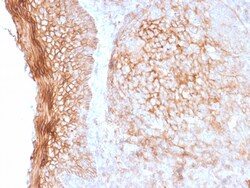
Biotium CD73 (Immuno-Oncology Target)(NT5E/2505), CF647 conjugate, 0.1mg/mL
CD73 (also designated ecto-5'-nucleotidase, E5NT, NT, NT5, NTE, eN and eNT) is a glycosyl-phosphatidylinositol (GPI)-anchored adhesion protein that catalyzes the dephosphorylation of extracellular purine and pyrimidine nucleotides to their corresponding bioactive nucleosides. CD73 is a dimer of two identical subunits that depends on GPI to link with the external face of the plasma membrane. Similar to other GPI-anchored proteins, CD73 mediates co-stimulatory signals in T cell activation. CD73 has few structural variants, yet elicits diverse biological function through differential regulation in endothelial cells (EC), subpopulations of B and T cells, germinal center follicular dendritic cells and on thymic medullary reticular fibroblasts. For example, IgG mediated neutralization of CD73 interferes with lymphocyte adhesion to EC, and blocks aggregation of germinal center B cells and follicular dendritic cells. Furthermore, IgG-mediated targeting of lymphocyte CD73, but not of
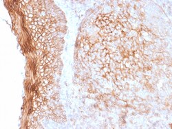
Biotium CD73 (Immuno-Oncology Target)(NT5E/2505), CF647 conjugate, 0.1mg/mL
CD73 (also designated ecto-5'-nucleotidase, E5NT, NT, NT5, NTE, eN and eNT) is a glycosyl-phosphatidylinositol (GPI)-anchored adhesion protein that catalyzes the dephosphorylation of extracellular purine and pyrimidine nucleotides to their corresponding bioactive nucleosides. CD73 is a dimer of two identical subunits that depends on GPI to link with the external face of the plasma membrane. Similar to other GPI-anchored proteins, CD73 mediates co-stimulatory signals in T cell activation. CD73 has few structural variants, yet elicits diverse biological function through differential regulation in endothelial cells (EC), subpopulations of B and T cells, germinal center follicular dendritic cells and on thymic medullary reticular fibroblasts. For example, IgG mediated neutralization of CD73 interferes with lymphocyte adhesion to EC, and blocks aggregation of germinal center B cells and follicular dendritic cells. Furthermore, IgG-mediated targeting of lymphocyte CD73, but not of
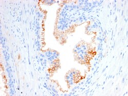
Biotium CD10 (Membrane Metalloendopeptidase)(MME/2580), CF647 conjugate, 0.1mg/mL
CD10 (also known as Common Acute Lymphocytic Leukemia Antigen (CALLA)) is a type II transmembrane protein. It is a cell surface enzyme with neutral metalloendopeptidase activity, which inactivates a variety of biologically active peptides. CD10 is expressed on the cells of lymphoblastic, Burkitt's, and follicular germinal center lymphomas, and on cells from patients with chronic myelocytic leukemia (CML). It is also expressed on the surface of normal early lymphoid progenitor cells, immature B cells within adult bone marrow and germinal center B cells within lymphoid tissue.Primary antibodies are available purified, or with a selection of fluorescent CF Dyes and other labels. CF Dyes offer exceptional brightness and photostability. Note: Conjugates of blue fluorescent dyes like CF405S and CF405M are not recommended for detecting low abundance targets, because blue dyes have lower fluorescence and can give higher non-specific background than other dye colors.
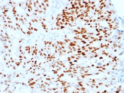
Biotium Estrogen Receptor, alpha (Marker of Estrogen Dependence)(ESR1/1904), CF647 conjugate, 0.1mg/mL
This monoclonal antibody is specific to estrogen receptor alpha (ER alpha) and shows minimal cross-reaction with other members of the family. ER is an important regulator of growth and differentiation in the mammary gland. Presence of ER in breast tumors indicates an increased likelihood of response to anti-estrogen (e. g. tamoxifen) therapy. It strongly stains nuclei of epithelial cells in breast carcinomas. Primary antibodies are available purified, or with a selection of fluorescent CF Dyes and other labels. CF Dyes offer exceptional brightness and photostability. Note: Conjugates of blue fluorescent dyes like CF405S and CF405M are not recommended for detecting low abundance targets, because blue dyes have lower fluorescence and can give higher non-specific background than other dye colors.
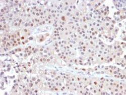
Biotium p21WAF1 (Tumor Suppressor Protein)(CIP1/2489R), CF647 conjugate, 0.1mg/mL
This MAb recognizes a 21 kDa protein, identified as the p21WAF1 tumor suppressor protein. This MAb is highly specific to p21 and shows no cross-reaction with other closely related mitotic inhibitors. p21WAF1 is a specific inhibitor of cdk's and a tumor suppressor involved in the pathogenesis of a variety of malignancies. The expression of this gene acts as an inhibitor of the cell cycle during G1 phase and is tightly controlled by the tumor suppressor protein p53. Its expression is induced by the wild type, but not mutant, p53 suppressor protein. Normal cells generally display a rather intense nuclear p21 expression. Loss of p21 expression has been reported in many carcinomas (gastric carcinoma, non-small cell lung carcinoma, thyroid carcinoma). Primary antibodies are available purified, or with a selection of fluorescent CF Dyes and other labels. CF Dyes offer exceptional brightness and photostability. Note: Conjugates of blue fluorescent dyes like CF405S and CF405M are
Biotium von Willebrand Factor / Factor VIII Related-Ag (Endothelial Marker) (VWF/1465), CF647 conjugate, 0.1mg/mL
von Willebrand Factor (vWF) is a multimeric glycoprotein that is found in endothelial cells, plasma and platelets. It acts as a carrier protein for Factor VIII and promotes platelet adhesion and aggregation. vWF undergoes a variety of posttranslational modifications that influence the affinity and availability for Factor VIII, including cleavage of the propeptide and formation of N-terminal disulfide bonds. This antibody helps to establish the endothelial nature of some lesions of disputed histogenesis, e.g. Kaposi's sarcoma and cardiac myxoma. It is widely used for differentiating vascular lesions from those of other tissue differentiation within a panel of other vascular markers although not all tumors of endothelial differentiation contain this antigen.Primary antibodies are available purified, or with a selection of fluorescent CF Dyes and other labels. CF Dyes offer exceptional brightness and photostability. Note: Conjugates of blue fluorescent dyes like CF405S and C
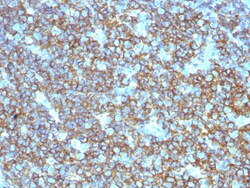
Biotium CD20 / MS4A1 (B-Cell Marker), CF647 conjugate, 0.1mg/mL
Recognizes a protein of 30-33 kDa, which is identified as CD20. It is a non-Ig differentiation antigen of B-cells and its expression is restricted to normal and neoplastic B-cells, being absent from all other leukocytes and tissues. CD20 is expressed by pre B-cells and persists during all stages of B-cell maturation but is lost upon terminal differentiation into plasma cells. This antibody can be used for immunophenotyping of leukemia and malignant cells, B lymphocyte detection in peripheral blood and B cell localization in tissues. It reacts with the majority of B-cells present in peripheral blood and lymphoid tissues and their derived lymphomas. In lymphoid tissue, germinal center blasts and B-immunoblasts are particularly reactive. It is a reliable antibody for ascribing a B-cell phenotype in known lymphoid tissues. Rarely, CD20-positive T-cell lymphomas have been reported. Reactivity has also been noted with Reed-Sternberg cells in cases of Hodgkin's disease, particularly
Biotium Cytokeratin, pan (Epithelial Marker), CF647 conjugate, 0.1mg/mL
Twenty human keratins are resolved with two-dimensional gel electrophoresis into acidic (pI 6.0) subfamilies. This antibody cocktail recognizes acidic (Type I or LMW) and basic (Type II or HMW) cytokeratins, which 67 kDa (CK1); 64 kDa (CK3); 59 kDa (CK4); 58 kDa (CK5); 56 kDa (CK6); 52 kDa (CK8); 56.5 kDa (CK10); 50 kDa (CK14); 50 kDa (CK15); 48 kDa (CK16); 40 kDa (CK19). Many studies have shown the usefulness of keratins as markers in cancer research and tumor diagnosis. It is a broad spectrum anti pan-cytokeratin antibody, which differentiates epithelial tumors from non-epithelial tumors e.g. squamous vs. adenocarcinoma of the lung, liver carcinoma, breast cancer, and esophageal cancer. It may be useful to characterize the source of various neoplasms and to study the distribution of cytokeratin containing cells in epithelia during normal development and during the development of epithelial neoplasms. This antibody stains cytokeratins present in normal and abnormal human tis
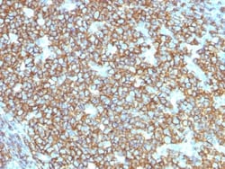
Biotium CD20 / MS4A1 (B-Cell Marker) (rIGEL/773), CF647 conjugate, 0.1mg/mL
This antibody recognizes a protein of 30-33 kDa, which is identified as CD20. It is a non-Ig differentiation antigen of B-cells and its expression is restricted to normal and neoplastic B-cells, being absent from all other leukocytes and tissues. CD20 is expressed by pre B-cells and persists during all stages of B-cell maturation but is lost upon terminal differentiation into plasma cells. This MAb can be used for immunophenotyping of leukemia and malignant cells, B lymphocyte detection in peripheral blood and B cell localization in tissues. It reacts with the majority of B-cells present in peripheral blood and lymphoid tissues and their derived lymphomas. In lymphoid tissue, germinal center blasts and B-immunoblasts are particularly reactive. It is a reliable antibody for ascribing a B-cell phenotype in known lymphoid tissues. Rarely, CD20-positive T-cell lymphomas have been reported. Reactivity has also been noted with Reed-Sternberg cells in cases of Hodgkin's disease, par
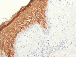
Biotium Cytokeratin 10 (Suprabasal Epithelial Marker)(KRT10/1948R), CF488A conjugate, 0.1mg/mL
This MAb recognizes a protein of 56. 5 kDa, identified as cytokeratin 10 (CK10). CK10 is expressed in all suprabasal layers of the epidermis. In the epidermis, expression of CK10 strictly parallels the extent of differentiation; it is absent in the basal layer, appears in the first suprabasal layers and increases in concentration towards the granular layer. However, CK10 is rarely detected in early stages of vulvar squamous carcinomas (tumors less than 2 cm, clinical stage I) regardless of the tumor grade. In larger and more advanced tumors (greater than 2 cm, clinical stages II and III), CK10 is detected very frequently. Expression of CK10 is related to maturation of malignant keratinocytes, being preferentially detected in more-differentiated parts. Primary antibodies are available purified, or with a selection of fluorescent CF Dyes and other labels. CF Dyes offer exceptional brightness and photostability. Note: Conjugates of blue fluorescent dyes like CF405S and CF40